
12
What Is AFib?
When the upper chambers quiver instead of squeezing — and why that can lead to a stroke.
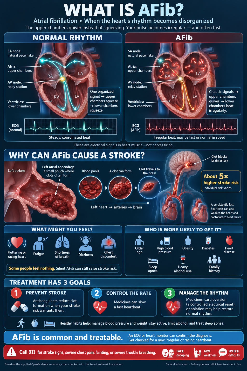
The heart has a natural pacemaker — the SA node — that fires an organized signal. The upper chambers (the atria) squeeze. That blood fills the lower chambers (the ventricles). The AV node relays the signal, the ventricles squeeze, and you get a pulse. Those are electrical signals in heart muscle, not nerves firing.
In atrial fibrillation that signal becomes disorganized. The atria quiver instead of squeezing. The ventricles still beat, but irregularly — and often fast. The pulse feels jumpy. The ECG looks like a messy, uneven line instead of a steady march.
The stroke risk is the part people underestimate. Because the left atrium is not emptying well, blood can pool in a small pouch called the left atrial appendage. A clot can form there, travel out the left heart into the arteries, and lodge in the brain. AFib raises stroke risk about five-fold; individual risk still varies. A persistently fast rate can also wear the heart out and contribute to heart failure.
What you might feel: a fluttering or racing heart, fatigue, shortness of breath, dizziness, or chest discomfort. Some people feel nothing. Silent AFib can still raise stroke risk. It is more common with older age, high blood pressure, obesity, diabetes, heart disease, sleep apnea, heavy alcohol use, and a family history.
Treatment has three jobs: prevent stroke (anticoagulants when your risk warrants them), control the rate if it is too fast, and manage the rhythm — medicines, a controlled electrical reset called cardioversion, or ablation in selected cases. Blood pressure, weight, activity, alcohol, and sleep apnea are the levers that actually help.
Get checked for a new irregular or racing heartbeat. Call 911 for stroke signs (face drooping, arm weakness, speech difficulty), severe chest pain, fainting, or severe trouble breathing.
 Watch on YouTube: Atrial Fibrillation Anatomy, ECG and Stroke, Animation.
Watch · Alila Medical Media
Atrial Fibrillation Anatomy, ECG and Stroke, Animation.
Three minutes: how the atria quiver instead of squeezing, what that looks like on an ECG, and how a clot from the left atrial appendage can travel to the brain.
Watch on YouTubeGeneral education only — not a substitute for advice from your healthcare provider. When in doubt, get examined.